

OCT & Retinal Photography in Picton for Early Detection
Eye care is essential for maintaining a healthy vision, and technological advancements have made it easier to detect potential problems early. Optical Coherence Tomography (OCT) and retinal photography are among the most essential innovations in eye health diagnostics. These procedures allow optometrists to capture detailed eye images, enabling them to identify underlying conditions that may not yet show visible symptoms. In Picton, these advanced techniques are making it possible to catch eye diseases early, helping to prevent severe vision loss. If you're looking for an Eye care clinic in Picton, these technologies are crucial in ensuring your vision remains clear and healthy.
What is OCT & Retinal Photography?
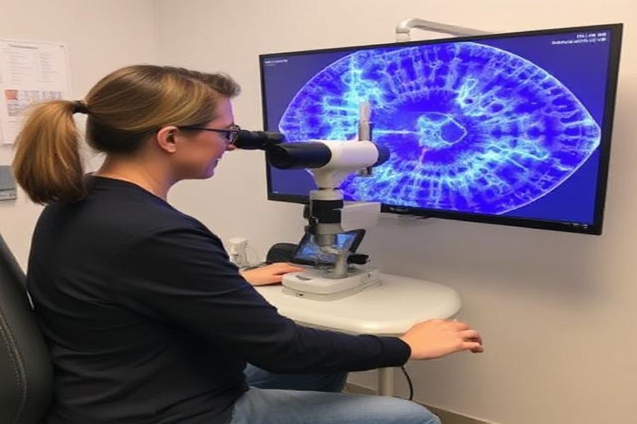
OCT (Optical Coherence Tomography) is a state-of-the-art imaging technique used by optometrists to capture detailed cross-sectional images of the retina. It works similarly to an ultrasound but uses light waves instead of sound waves. This allows for the visualization of the layers of the retina, giving optometrists a clear view of your eye’s internal structure.
Retinal Photography is another imaging technique that captures high-resolution photographs of the retina. These images provide a detailed view of the retina’s blood vessels, optic disc, and macula. Retinal photography is particularly effective at documenting changes over time, making it a valuable tool for monitoring eye conditions and assessing the progression of certain diseases.
Why is Early Detection Important?
Early detection of eye diseases is crucial for preserving vision. Many severe eye conditions, such as glaucoma, diabetic retinopathy, and macular degeneration, develop gradually, and their symptoms may not be noticeable in the early stages. Without routine eye exams and advanced diagnostic tools, individuals may not realize something is wrong until the damage is done.
OCT and retinal photography allow optometrists to spot problems at their earliest stages. This enables timely intervention, significantly reducing the risk of irreversible vision damage. Some of the conditions these technologies can help detect include:
- Glaucoma: A condition that damages the optic nerve, often caused by high eye pressure. OCT can measure the thickness of the optic nerve to detect early signs of damage.
- Diabetic Retinopathy: This is a problem caused by diabetes that affects the blood vessels in the retina. Retinal photography can identify abnormal blood vessels and leakage, which may indicate this condition.
- Age-related Macular Degeneration (AMD): A condition that affects the central part of the retina and can lead to blurred or distorted central vision. OCT allows optometrists to examine the macula in great detail, helping to detect early signs of AMD.
- Retinal Vein Occlusion (RVO): A blockage of the veins in the retina can lead to vision loss. Retinal photography can capture changes in the retinal blood vessels, making diagnosing it easier.
Benefits of OCT & Retinal Photography
- Non-invasive and Painless: Both OCT and retinal photography are non-invasive procedures that do not require any injections or incisions. They are pain-free and only take a few minutes to complete.
- Early Detection: These imaging techniques allow optometrists to detect eye diseases in their earliest stages, often before symptoms appear. Early treatment can prevent or slow the progression of many conditions.
- Improved Diagnosis: By providing a detailed view of the eye’s internal structures, OCT and retinal photography help optometrists make more accurate diagnoses. This ensures that you receive the proper treatment at the right time.
- Monitor Progression: Retinal photography helps track the progression of eye conditions over time, allowing optometrists to adjust your treatment plan as needed.
- Better Outcomes: Early diagnosis and treatment often improve outcomes, including preserving vision. Proper care can effectively manage many eye diseases, helping you maintain clear and healthy vision.
How Often Should You Have OCT & Retinal Photography?
While OCT and retinal photography can be extremely useful in detecting eye diseases, not everyone must undergo these tests yearly. The frequency of these procedures depends on a variety of factors, including:
- Your age: People over 40 are at higher risk for age-related eye conditions and should have regular eye exams to monitor for changes.
- Your Health: Diabetes, hypertension, and high cholesterol can increase your risk for eye diseases. Regular eye exams and imaging tests are essential if you have any of these conditions.
- Family history: If you have a family history of eye diseases such as glaucoma or macular degeneration, you may need more frequent monitoring.
- Symptom development: If you notice changes in your vision, such as blurriness, blind spots, or flashes of light, you should schedule an eye exam as soon as possible.
Conclusion
Incorporating OCT and retinal photography into routine eye exams is a game-changer in the early detection of eye diseases. These non-invasive imaging techniques allow optometrists in Picton to detect problems long before they cause noticeable symptoms, ensuring that treatment can begin at the earliest possible stage. With advancements in technology, maintaining eye health has never been more accessible, and these diagnostic tools can play a vital role in preserving your vision for years to come.
Regular eye exams are a critical part of overall health, so if you haven’t had an eye exam recently, it’s time to book one and take advantage of these modern diagnostic technologies. Early detection could make all the difference in maintaining your vision.